Bonapriso, rue Koloko,
Douala
doualaeyecenter@gmail.com
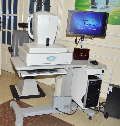
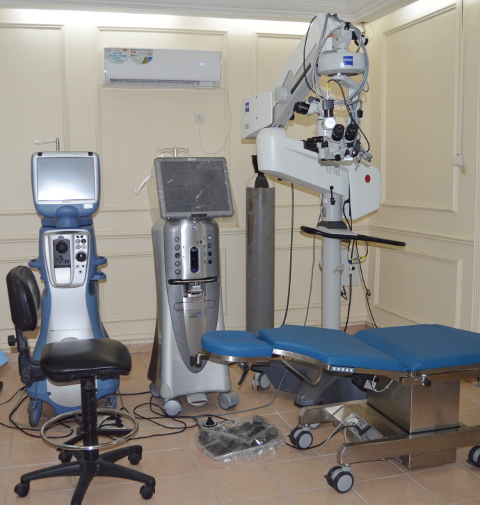
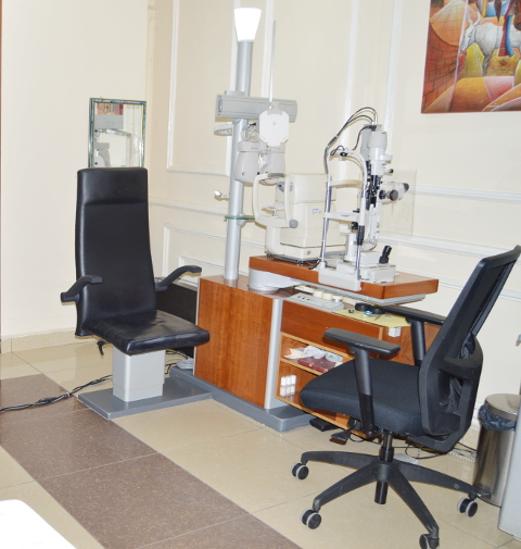
Advanced Technology
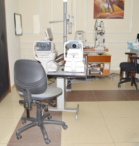
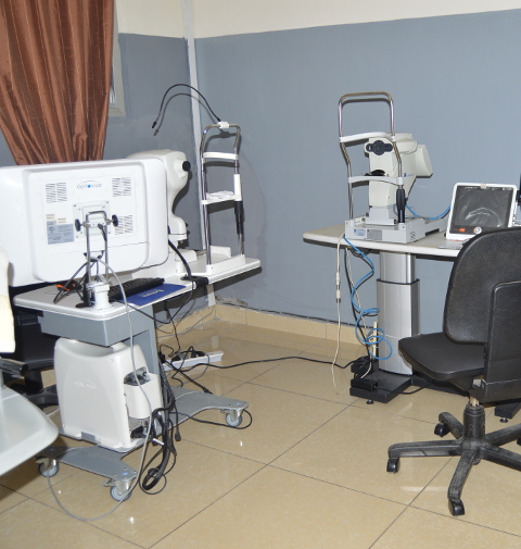
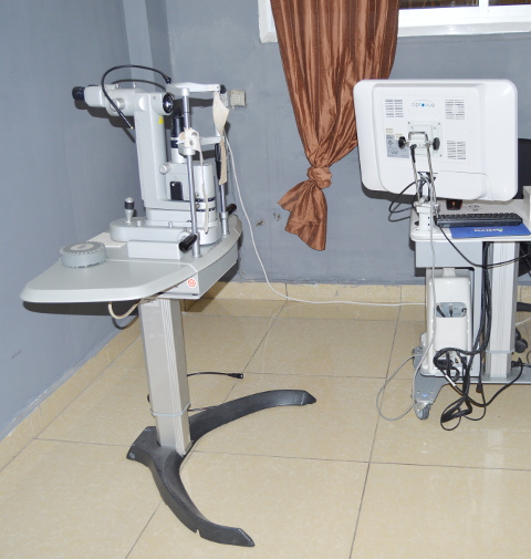
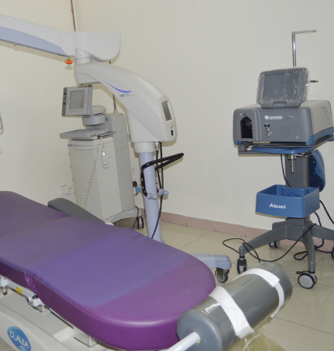
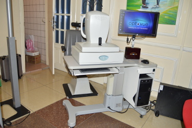
The Center is equipped with a complete technical platform of the latest generation.
Cares
For each visual problem, there is a suitable solution.
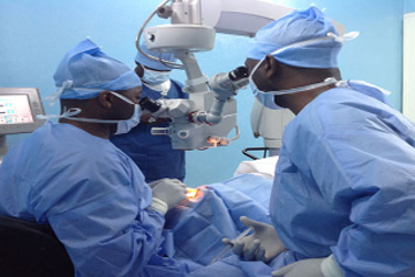
Douala International Eye Center is an ophthalmology center specializing in the prevention, medical and surgical treatment of eye diseases including surgery for myopia, hyperopia, astigmatism, presbyopia and cataract surgeries, cornea and keratoconus. Our center specializes in eye surgery, general ophthalmology. Below are some of the eye diseases:
PRK or REFRACTIVE PHOTOKERATEC TOMIE
The center has a state-of-the-art and complete technical platform for the diagnosis and monitoring of most eye pathologies.
This is the first technique using the Excimer Laser for the treatment of vision defects.
This surgical technique involves three operating steps:
1- Mechanical peeling of the corneal epithelium, with a brush or scarifier, with or without diluted alcohol.
2- Application of the laser beam, computer-assisted, on the cornea underlying the epithelium.
3- Application of mitomycin diluted 0.02% in case of deep ablation to avoid subepithelial scarring (Haze).
The abrasion of the superficial cells of the cornea causes postoperative pain for a few days requiring treatment with painkillers.
Visual recovery is slow; one to two weeks.
This technique is reserved for medium degrees of vision defects: myopia, hyperopia, astigmatism.
Both eyes are operated separately, or simultaneously.
The Excimer Laser is used in various refractive surgery techniques.
TRANS PRK
Technique specific to the Schwind Amaris Excimer laser:
- Abrasion of surface epithelial cells by the laser
- Followed by collagen abrasion by the same Excimer laser.
The advantages of this technique are:
- Speed: processing in a few seconds
- Proximity of epithelial ablation and collagen ablation favoring faster healing and reduction of the postoperative painful period
- This technique avoids mechanical ablation and the use of diluted alcohol on the epithelium.
- This is the only surgical technique without any contact with the cornea, the laser itself doing all the steps of the surgery.
TransPRK:
The only treatment by which, without contact with the eye, and in a more precise way than before, the epithelium is ablated by the excimer laser and then the collagen is ablated for the correction of refractive ametropia.
TransPRK – “No contact” treatment:
The ablation of the epithelium and the refractive anomaly are carried out immediately one after the other. As a result, the total time of the intervention is significantly reduced.
The Excimer Laser is used in different refractive surgery techniques
LASIK
This is the technique most frequently used by refractive surgeons in the United States and Europe. Available in our center located in Brussels (Belgium):
- Lasik or Laser In Situ Keratomileusis begins with the production of a thin superficial corneal lamella (hood).
- This flap is produced using an automated microkeratome or a Femtosecond laser.
- It is then lifted and remains attached to part of the corneal periphery
- The computer-assisted Excimer laser beam is applied to the underlying part.
The flap is then repositioned, without sutures. Adherence occurs spontaneously within a few days.
The postoperative course is painless and visual recovery is rapid, in a few hours, because no abrasion is made on the surface of the eye.
Lasik is aimed at medium and strong degrees of vision defects: myopia, hyperopia, astigmatism.
Since 2007, he has been treating presbyopia associated with myopia, hyperopia and astigmatism using the Optimized Global Monovision technique, which promotes a better accommodation margin for near vision.
Since 2008, Doctor Assaf has also been performing corneal flaps using the Femtosecond LDV laser: corneas that are too arched or too flat, corneas that are too thin, etc.
He also uses the latest software to treat corneas by refractive surgery while preserving a natural prolate shape of the cornea, optimizing visual quality (Eye Q).
Since 2013, Doctor Assaf has been using the Lasik micromonovision technique with the Schwind Amaris laser for the treatment of presbyopia associated with myopia, hyperopia and astigmatism.
The Excimer Laser is used in different refractive surgery techniques
TRAITEMENT PERPERSONALIZED TREATMENT BY ABERROMETRY OR WAVE FRONT WAVE FRONT GUIDED CUSTOM LASER
Like our fingerprints, our eyes have an individual shape with their own characteristics. The personalized treatment by aberrometry uses software allowing the treatment of optical aberrations specific to each eye.
Its objective is to improve visual quality compared to glasses, contact lenses or conventional treatment.
Until 2002, electronic diagnostic devices could detect myopia, hyperopia and astigmatism. These optical defects account for approximately 90% of total eye abnormalities. The remaining 10% are called higher order optical aberrations.
Thanks to new devices called aberrometers, we can currently measure and treat these higher-order aberrations, in a personalized way, with respect to each patient.
Analysis by Wave Front:
It is a technique originally developed by astrophysicists and which is currently applied for the treatment of refractive errors.
The analysis of the reflection image through an optical grid makes it possible to analyze the optical aberrations characteristic of each patient.
- This analysis is done without contact with the eye by simply taking photos, which allow the analysis of the entire optical system of the eye, from the retina to the cornea.
- The measurement of aberrations is combined with the analysis of the anterior and posterior corneal topography, by a topographic map (Orbscan).
- The parameters measured by the Aberrometer and the Orbscan, are combined on a diskette which allows computer-assisted treatment of conventional anomalies (myopia, astigmatism) and higher order optical aberrations, during a PRK procedure, or LASIK.
The advantages of a personalized treatment by Aberrometry and Topoguided Corneal Wavefront are as follows:
- Personalized treatment according to the aberrations of each eye.
- Improvement of the best postoperative visual acuity, sometimes going to a view greater than 10/10.
- Improved night and sometimes daytime visual quality.
- Lower risk of reduction in best corrected visual acuity.
- Better stability and reduced risk of reprocessing.
- Possibility of treatment of severe ametropia not treatable until now by the conventional technique, although associated with a cornea of average thickness, or associated with a large pupillary diameter in the penumbra.
- Possibility of correction by reprocessing of patients previously operated by refractive surgery presenting irregular astigmatism, a small optical zone… resulting in nocturnal visual disturbances: Halos, dazzling by light…
It is important to note that the treatment by aberrometry will be mainly indicated in patients who will have, according to the preoperative results, significant aberrations or a large pupillary diameter in the penumbra and a favorable profile advocating a better result than with the conventional technique.
The Excimer Laser is used in different refractive surgery techniques
INTRAOCULAR IMPLANTS OF THE PHAQUE
Definition: this is the placement of an intraocular implant, correcting the refractive error, without removing the lens.
Indications: all refractive anomalies not operable by Excimer laser, namely:
High myopia > -10 diopters
High hypermetropia > +5 diopters
High astigmatism > 5 diopters
Cornea too thin
Types of implants
ARTISAN
ARTIFLEX
VISIAN ICL
BIOPTICS
KR: RADIARY KERATOTOMY (no longer practiced)
This is the first surgical technique used for the correction of myopia.
This technique was commonly used in the 70s and 80s, before the arrival of the Excimer Laser.
It consists of making several radial corneal incisions, from the periphery to the center.
The number and length of incisions depends on the degree of myopia.
The purpose of this intervention is the flattening of the anterior cornea allowing the retraction of the focal point of the images on the retina.
The incisions are made using a pre-calibrated diamond knife.
Radial keratotomy treats low myopia and low astigmatism.
ARTIFLEX
It is a myopic phakic implant (without removal of the natural lens) with a specific characteristic of being foldable.
Its introduction is carried out through a small incision of 3 mm.
Its fixation is like the Artisan Irienne.
Its advantage is to reduce the size of the incision and reduce the incidence of postoperative astigmatism.
Its disadvantage is that it predisposes to a more frequent deposit of pigments given the material used and requiring a longer anti-inflammatory treatment than the Artisan.
Visual recovery with this implant is obtained the day after the intervention.
Reversibility = the Artiflex implant is easy to remove if an exchange is necessary.
It also corrects high myopia up to -15.5 diopters.
ARTIFLEX Toric: Corrects astigmatism associated with myopia simultaneously.
Types of implants
ARTISAN
ARTIFLEX
VISIAN ICL
BIOPTICS
INDICATIONS OF THE DIFFERENT REFRACTIVE SURGERY TECHNIQUES
INDICATIONS OF THE DIFFERENT TECHNIQUES
Radial keratotomy: Myopia -1 to -3 diopters
PRK or Lasek: Myopia -1 to -3 diopters, astigmatism -1 to -3 diopters
TRANS PRK: Myopia, hypermetropia and Astigmatism from +/-1 to +/-5 diopters
Lasik and Femtolasik:
Myopia: -1 to -10 diopters
Astigmatism: +/- 1 to +/- 5 diopters
Hyperopia: + 1 to + 5 diopters
Aberrometry: Personalized treatment by Aberrometry and Ultra Thin Lasik:
Myopia: -1 to -10 diopters
Astigmatism: -1 to -6 diopters
Presence of significant optical aberrations
Large pupil diameter in the dark
Indication for improvement in visual quality
Thin cornea
Special features of all surgical techniques
KR, PRK, Lasek, Trans PRK, Lasik and Femtolasik are techniques that can lead to regression, under or over postoperative correction due to the individual variability of human corneal tissue, compared to the ablation profile.
The regression rate is 1 to 2%. A free touch-up is then necessary 2 to 3 months after the first intervention.
Retouching for over or under correction after radial keratotomy or PRK can be performed by PRK or Lasik
Alterations after (Femto)lasik will only be done by (Femto)lasik
Inclusion criteria for refractive surgery
Age: > 19 years old
Myopia, astigmatism or hypermetropia stable for more than 2 years
Absence of eye diseases: keratoconus, progressive glaucoma,
chronic herpes...
Absence of chronic autoimmune diseases and collagenoses: lupus, periarteritis nodosa, scleroderma…
Absence of poorly controlled ocular diabetes
Good motivation: professional, esthétique, sportive
COMPLICATIONS OF REFRACTIVE SURGERY
Complications specific to each surgical technique:
Radial keratotomy
- Daily visual fluctuations
- Night glare and halos
- Secondary or progressive hyperopia
- Theoretical weakening of the globe
PRK–Trans PRK–Lasek
- Postoperative pain 2 to 3 days
- Delay of epithelialization
- Haze or subepithelial scarring, responsible for visual haze, gradually diminishing: 1 to 2%
- Nocturnal halos, in case of high myopia, and large pupillary diameter and in case of subepithelial scarring.
- Low risk of postoperative infection or inflammation: 1/4000
Lasik–Femtolasik
- Risk of displacement or folds of the corneal flap in the first days in the event of brutal friction or folds of the flap, hence the need to wear a rigid shell, at night, for 2 weeks after the intervention.
- Risk of dry eye requiring the instillation of artificial tears, one month after the operation.
- Risk of invasion of the space under the corneal flap by epithelial cells, requiring rinsing (0.1%).
- Risk of an inflammatory reaction under the corneal flap requiring anti-inflammatory treatment or rinsing (0.5%).
- Risk of nocturnal halos, in case of high myopia and larger pupillary diameter
- Low risk of postoperative infection or inflammation: 1/5000
- Risk of ectasia or irregular astigmatism: This is an exceptional and late complication, occurring mainly on corneas with frustrated keratoconus or other corneal pathologies not diagnosed or not apparent preoperatively. This complication occurs especially when the flaps are thick and the ablation of the corneal tissue is deep.
Aberrometer
- Personalized treatment by aberrometry, either by Lasik or PRK.
- Complications identical to the chosen technique
- Less frequency of halos and nocturnal glare.
S.M.I.L.E (SMall Incision Lenticule Extraction)
This is a new surgical technique for myopia and astigmatism by Femto Second laser. It is performed under local anesthesia by drops.
The laser pre-cuts a small lenticule in the thickness of the cornea corresponding to the volume of myopia and astigmatism to be treated.
The laser makes a small peripheral incision (2 to 4 mm wide) through which the surgeon will dissect the lenticule and extract it from the cornea.
The results of this technique in the case of myopia and astigmatism are predictable and lead to very good refractive results.
ADVANTAGES
Absence of creation of a fragile flap in the event of friction and trauma.
- Less dryness
DISADVANTAGES
- Slower visual recovery.
- Optimal vision after a few days and not the next day like Lasik.
- Need for longer-lasting anti-inflammatory treatment.
- SMILE, unlike Lasik, does not currently treat strong astigmatism and farsightedness.
- Difficulty of reprocessing in case of over and under correction.
COMPLICATIONS
The complications are identical to refractive surgeries by Laser (infections, inflammation, night vision disorders, etc.) except for the folds and displacement of the flap.
The Excimer Laser is used in various refractive surgery techniques:
PRK
TRANSPRK
Lasek
LASIK
Femtolasik
Aberrometry and personalized treatment by wavefront
ULTRA-THIN LASIK and Sub-bowman Keratomileusis (SBKM)
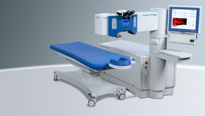
LASER Excimer
The Excimer Laser whose name comes from EXCIted diMER uses the energy of the photons released by the electrical excitation of the Dimer Argon Fluor, to carry out a superficial abrasion of the cornea and correct the refractive defects: myopia, hyperopia, astigmatism.
The Excimer laser treatment is always computer assisted to define the profile of the corneal ablation according to the preparatory examination.
ARTISAN
Preferably local or general anesthesia.
Opening a small corneal incision.
Placement of a lens that is attached to the iris (ARTISAN)
Corneal suture at the end of the intervention to be removed a few weeks after the intervention.
Advantage of the implant, Artisan type:
Clinical experience since the 80s.
5 mm myopic implant, which can correct up to -23 diopters.
6 mm myopic implant, which can correct up to -16 diopters.
Hypermetropic implant of 5 mm, which can correct up to +12 diopters.
Little risk of inducing a cataract.
Little or no risk of corneal decompensation.
Reversible treatment
CRAFTSMAN Toric:
Implant correcting astigmatism up to 7.5 diopters, simultaneously with high myopia
Possible complications:
Risk of pigment dispersion or ocular hypertension, controllable by local treatment with eye drops.
Low risk of infection and postoperative inflammation: 2 to 3/1000
Exceptional risk of decreased intraocular cell density requiring removal of the implant
BIOPTICS
This is an operative technique combining the placement of an Artisan, Artiflex or Visian ICL intraocular implant followed by treatment with Excimer Laser PRK, or Lasik.
Indication: strong ametropia, or refractive anomaly, which cannot be treated, only by Lasik or by implant.
Examples:
-20 diopter myopic, with -5 diopter astigmatism
Treatment with Artisan 6 mm implant or Visian ICL of -18 diopters and
-3 diopries of astigmatism
Supplemented by Lasik for residual myopia of -2 diopters and residual astigmatism of -2 diopters
Hyperopic +8 diopters, with an astigmatism of +5 diopters
Placement of an Artisan or Visian ICL implant of +5 diopters with astigmatism of +3 diopters
Complementary Lasik treatment for hyperopia of +2 diopters and astigmatism of +2 diopters.
Types of implants
ARTISAN
ARTIFLEX
VISIAN ICL
BIOPTICS
ULTRA-THIN LASIK
This is the Lasik technique with a very thin flap (90 to 110µ, average of 100µ) allowing laser ablation directly under Bowman's membrane. The thin flap can be produced either by microkeratome designed for this purpose, or by Femtosecond laser.
This technique has the following clinical advantages:
- Economy of corneal tissue
- Extension of indications to higher myopia and astigmatism
- Maintenance of corneal architecture and stability
- Reduction of corneal weakening by leaving a posterior stroma with a thickness greater than 300µ
- Reprocessing of residual myopia easier by saving tissue
The Excimer Laser is used in various refractive surgery techniques:
PRK
TRANSPRK
Lasek
LASIK
Femtolasik
Aberrometry and personalized treatment by wavefront
ULTRA-THIN LASIK and Sub-bowman Keratomileusis (SBKM)
FEMTOLASIK by FEMTOSECOND LASER LDV Z6 (ZIEMER)
The Femtosecond laser is a surgical tool intended for cutting the cornea.
Doctor Assaf uses the Ziemer LDV Z6 femtosecond.
Its applications are:
- Refractive: Lasik surgery for myopia, hyperopia, astigmatism, presbyopia (corneal tunnel and corneal inlay)
- Therapeutic: lamellar corneal transplant, realization of corneal tunnels for the establishment of intra-corneal rings for the treatment of keratoconus.
Other clinical indications are under study. This is a technology approved in the United States (FDA approval) whose studies date back to the early 1980s and marketing in the early 1990s.
The Femtosecond Laser works with extremely short pulses, of the order of a Femto second, i.e. 10-15 seconds. This low pulse duration avoids thermal effects. It allows, thanks to an effect on the electrons of the atoms, the creation of several thousand small bubbles of gas. The lifting of the flap is then possible by the surgeon thanks to a forceps which crosses the plan of action of the laser at the level of the microbubbles.
Femtosecond Laser cutting makes it possible to predetermine the diameter of the cover, the thickness of the cover and the length of the hinge. Like the microkeratome, it requires the placement of a suction ring outside the cut.
Its indications are preferred in case of a cornea that is too arched or too flat.
FEMTOSECOND versus Microkeratome: The refractive result following Lasik performed both by Microkeratome and by Femtosecond is not significantly different. To date, no clinical study has demonstrated the superiority of one method over the other. Nevertheless, from the personal experience of Doctor Assaf, the Femtosecond could have privileged indications in corneas that are too flat or too arched, and small palpebral deviations, and the Microkeratome in the event of hyperopia to achieve an off-centered flap because the axis vision of farsighted people is always off-centre. It is easier to make an off-center flap by Microkeratome than by femtosecond.
The microkeratome is also indicated in the realization of flap after radial keratotomy.
The major drawback of the Lasik by Femtosecond technique is its cost which is relatively higher (25% more expensive than Lasik by Microkeratome) because this technology uses expensive disposable equipment and the price of the Femtosecond laser is very high. Its other disadvantage is the longer duration of the suction and the realization of the hood. As a result, according to some studies, it would induce corneal dryness more frequently.
(www.ziemergroup.com/)